CAZypedia needs your help! We have many unassigned GH, PL, CE, AA, GT, and CBM pages in need of Authors and Responsible Curators.
Scientists at all career stages, including students, are welcome to contribute to CAZypedia. Read more here, and in the 10th anniversary article in Glycobiology.
New to the CAZy classification? Read this first.
*
Consider attending the 15th Carbohydrate Bioengineering Meeting in Ghent, 5-8 May 2024.
Glycoside Hydrolase Family 172
This page has been approved by the Responsible Curator as essentially complete. CAZypedia is a living document, so further improvement of this page is still possible. If you would like to suggest an addition or correction, please contact the page's Responsible Curator directly by e-mail.
| Glycoside Hydrolase Family GH172 | |
| Clan | None |
| Mechanism | retaining |
| Active site residues | known |
| CAZy DB link | |
| http://www.cazy.org/GH172.html | |
Substrate specificities
Glycoside hydrolase family 172 (GH172) includes α-D-arabinofuranosidases and α-D-fructofuranosidases. This family was established following the discovery of αFFase1 from Bifidobacterium dentium by Kashima et al. in 2021 [1]. αFFase1 hydrolyzes the alkylated glycosides Me-α-D-Araf and Me-α-D-Fruf. In nature, it catalyzes the dehydrating condensation reaction of inulobiose (β-D-Fruf-(2→1)-α-D-Fruf) to difructose dianhydride I (DFA I, α-D-Fruf-1,2′:2,1′-β-D-Fruf). The dehydrating condensation reaction reaches equilibrium when the ratio of DFA and inulobiose is 9:1. αFFase1 is less specific for D-Fru at the non-reducing end and is able to catalyze the dehydrating condensation of β-D-Frup-(2→1)-α-D-Fruf to diheterolevulosan II (DHL II, α-D-Frup-1,2′:2,1′-β-D-Fruf). Physiologically, it is believed that after degradation of DFA I to inulobiose, inulobiose is degraded to D-Fru by GH32 β-D-fructofuranosidase, and then the produced monosaccharides are metabolized by the microorganism. DFA I is an oligosaccharide found in caramel, and since the degradation system of DFA I by bifidobacteria has been clarified, DFA I has attracted a certain attention in the food industry.
Also, some GH172 enzymes which physiologically functions as α-D-arabinofuranosidase, was reported in 2023 by Al-Jourani et al. (DgGH172a, DgGH172b, DgGH172c [2]) and Shimokawa et al. (ExoMA1 [3]). In particular, ExoMA1 was compared in detail with αFFase1, and it was found that its α-D-fructofuranosidase activity is extremely weak. These enzymes are believed to be involved in the degradation system of D-arabinan in the cell walls of Mycobacteria and other acid-fast bacteria, and are expected to be applied to the development of therapeutic, preventive, and diagnostic agents for infectious diseases.
Kinetics and Mechanism
As of 2023, all of the enzymes reported in GH172 catalyze reactions by an anomer-retaining mechanism. When pNP-D-Araf is hydrolyzed by αFFase1 [1] or ExoMA1 [3], the initial product was identified by 1H NMR to have the same anomeric conformation as the substrate. In addition, when pNP-α-D-Araf was enzymatically treated in the presence of organic solvents, transglycosylation products were detected by TLC. Enzyme kinetic experiments with this substrate were first reported for αFFase1, with Km and kcat values of 2.71 mM and 127.5 s-1, respectively; kinetic constants for ExoMA1 were comparable.
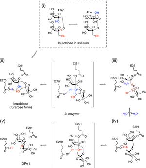
The dehydrating condensation reaction of inulobiose to DFA I by αFFase1 was proposed to proceed as follows (Figure 1).
(i) The reducing end sugar of inulobiose changes its furanose/pyranose and α/β forms in the internal cavity of αFFase1 as well as in solution because of mutarotation.
(ii) The active site of αFFase1 selectively accommodates the α-furanose form in the −1 subsite. The +1 subsite can also accommodate the pyranose moiety of Frupβ2,1Fru. Glu270 donates a proton (general acid catalysis) to the O2 hydroxy group of α-Fruf at the −1 subsite, and Glu291 works as a nucleophile to the anomeric carbon (C2). After this step, the O2 hydroxy group is released from the substrate as a water molecule.
(iii) Rotations of the glycosidic bond and the C1–C2 bond of the +1-subsite sugar are required for the intramolecular transfer reaction to occur.
(iv) When the C1 hydroxy group of fructose at the +1 subsite is appropriately positioned for proton acceptance (general base catalysis) by Glu270, deglycosylation of Glu291 is facilitated.
(v) After the reaction at the active site, DFA I is released through the channel to the inner cavity of the hexamer of αFFase1.
The opposite reaction from DFA I (v) to inulobiose (i) is expected to be similar to the standard retaining reaction mechanism of GHs. The kcat/Km were 0.813 mM-1·s-1 and 0.0378 mM-1·s-1 for inulobiose dehydration and DFA I hydrolysis, respectively.
Catalytic Residues
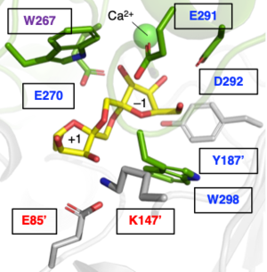
X-ray crystallographic analysis of the catalytic residues of αFFase1 [1] and ExoMA1 [3] revealed that the hydrolysis and dehydrating condensation reactions reported in GH172 are catalyzed by two glutamate residues, which act as the catalytic nucleophile and the general acid/base. In particular, site-directed mutagenesis of the two catalytic residues of αFFase1 and the amino acid residues that form the −1 and +1 subsites was performed, and the mode of ligand recognition and catalytic mechanism has been analyzed in detail (Figure 2).
Three-dimensional structures
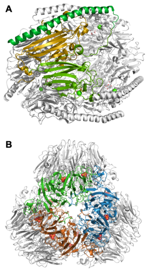
GH172 has a double jelly-roll (DJR) fold consisting of two β-jelly roll domains in the monomer. This is a fold that had not been reported in CAZymes prior to the establishment of GH172. The basic structure is a C3 symmetrical trimer, and the active site is formed by the first β-jelly roll and the second β-jelly roll of the adjacent subunit. The addition of α-helix or loops to the basic structure enables the elaboration of a higher quaternary structure. For example, in αFFase1, a long C-terminal α-helix covers the outside of the overall structure to maintain a D3 dihedral hexameric structure (Figure 3A [1]), while in ExoMA1, loops in the DJR are elongated to form a tetrahedral dodecamer (Figure 3B [3]). A small molecule predicted to be a phosphate is coordinated between the trimers in ExoMA1. In addition, BACUNI_00161, a protein of unknown function which belongs to GH172, forms a hexamer by hooking the C-terminal α-helix between the subunits facing each other. It is possible that various other oligomeric forms exist, suggesting that GH172 is a structurally diverse family.
Family Firsts
- First stereochemistry determination
- DFA I synthase/hydrolase αFFase1 from Bifidobacterium dentium JCM 1195 1H NMR.
- First catalytic nucleophile identification
- DFA I synthase/hydrolase αFFase1 from Bifidobacterium dentium JCM 1195 by X-ray crystallography and site-directed mutagenesis.
- First general acid/base residue identification
- DFA I synthase/hydrolase αFFase1 from Bifidobacterium dentium JCM 1195 by X-ray crystallography and site-directed mutagenesis.
- First 3-D structure
- BACUNI_00161 from Bacteroides uniformis ATCC 8492 by X-ray crystallography. This structure was determined as part of structural genomics, and therefore the function of this enzyme has not been confirmed, although it is expected to be α-D-arabinofuranosidase based on sequence homology. The next one determined was DFA I synthase/hydrolase αFFase1 from Bifidobacterium dentium JCM 1195 by X-ray crystallography..
References
- Kashima T, Okumura K, Ishiwata A, Kaieda M, Terada T, Arakawa T, Yamada C, Shimizu K, Tanaka K, Kitaoka M, Ito Y, Fujita K, and Fushinobu S. (2021). Identification of difructose dianhydride I synthase/hydrolase from an oral bacterium establishes a novel glycoside hydrolase family. J Biol Chem. 2021;297(5):101324. DOI:10.1016/j.jbc.2021.101324 |
- Al-Jourani O, Benedict ST, Ross J, Layton AJ, van der Peet P, Marando VM, Bailey NP, Heunis T, Manion J, Mensitieri F, Franklin A, Abellon-Ruiz J, Oram SL, Parsons L, Cartmell A, Wright GSA, Baslé A, Trost M, Henrissat B, Munoz-Munoz J, Hirt RP, Kiessling LL, Lovering AL, Williams SJ, Lowe EC, and Moynihan PJ. (2023). Identification of D-arabinan-degrading enzymes in mycobacteria. Nat Commun. 2023;14(1):2233. DOI:10.1038/s41467-023-37839-5 |
-
Shimokawa, M., Ishiwata, A., Kashima, T. et al. (2023) Identification and characterization of endo-α-, exo-α-, and exo-β-D-arabinofuranosidases degrading lipoarabinomannan and arabinogalactan of mycobacteria. Nature Communications 14, 5803. DOI:10.1038/s41467-023-41431-2


