CAZypedia needs your help!
We have many unassigned pages in need of Authors and Responsible Curators. See a page that's out-of-date needs a touch-up? - you are welcome to become a CAZypedian. Here's how.
Scientists at all career stages, including students, are welcome to contribute
Read more about CAZypedia here, and in the 10th anniversary article in Glycobiology.
New to the CAZy classification? Read this first.
Difference between revisions of "File:CE3 Figure.png"
Joel Weadge (talk | contribs) (Crystal structure of ''Tc''AE206 from ''Talaromyces cellulolyticus'' (PDB ID 5B5S). Colours correspond to α-helices (cyan), β-sheets (magenta), loops (wheat), disulfide bond (yellow), calcium ion (orange) and the active site residues (green).) |
(No difference)
|
Latest revision as of 10:54, 2 June 2020
Crystal structure of TcAE206 from Talaromyces cellulolyticus (PDB ID 5B5S). Colours correspond to α-helices (cyan), β-sheets (magenta), loops (wheat), disulfide bond (yellow), calcium ion (orange) and the active site residues (green).
File history
Click on a date/time to view the file as it appeared at that time.
| Date/Time | Thumbnail | Dimensions | User | Comment | |
|---|---|---|---|---|---|
| current | 10:54, 2 June 2020 | 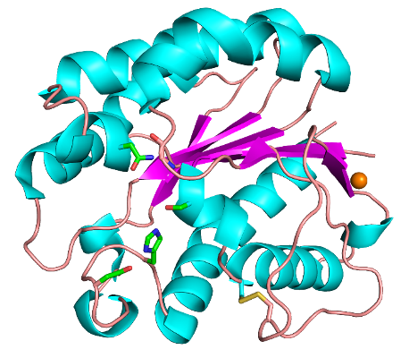 | 458 × 400 (152 KB) | Joel Weadge (talk | contribs) | Crystal structure of ''Tc''AE206 from ''Talaromyces cellulolyticus'' (PDB ID 5B5S). Colours correspond to α-helices (cyan), β-sheets (magenta), loops (wheat), disulfide bond (yellow), calcium ion (orange) and the active site residues (green). |
You cannot overwrite this file.
File usage
The following page uses this file: