CAZypedia needs your help! We have many unassigned GH, PL, CE, AA, GT, and CBM pages in need of Authors and Responsible Curators.
Scientists at all career stages, including students, are welcome to contribute to CAZypedia. Read more here, and in the 10th anniversary article in Glycobiology.
New to the CAZy classification? Read this first.
*
Consider attending the 15th Carbohydrate Bioengineering Meeting in Ghent, 5-8 May 2024.
File:Fig.2 300.png
Original file (1,653 × 991 pixels, file size: 1.74 MB, MIME type: image/png)
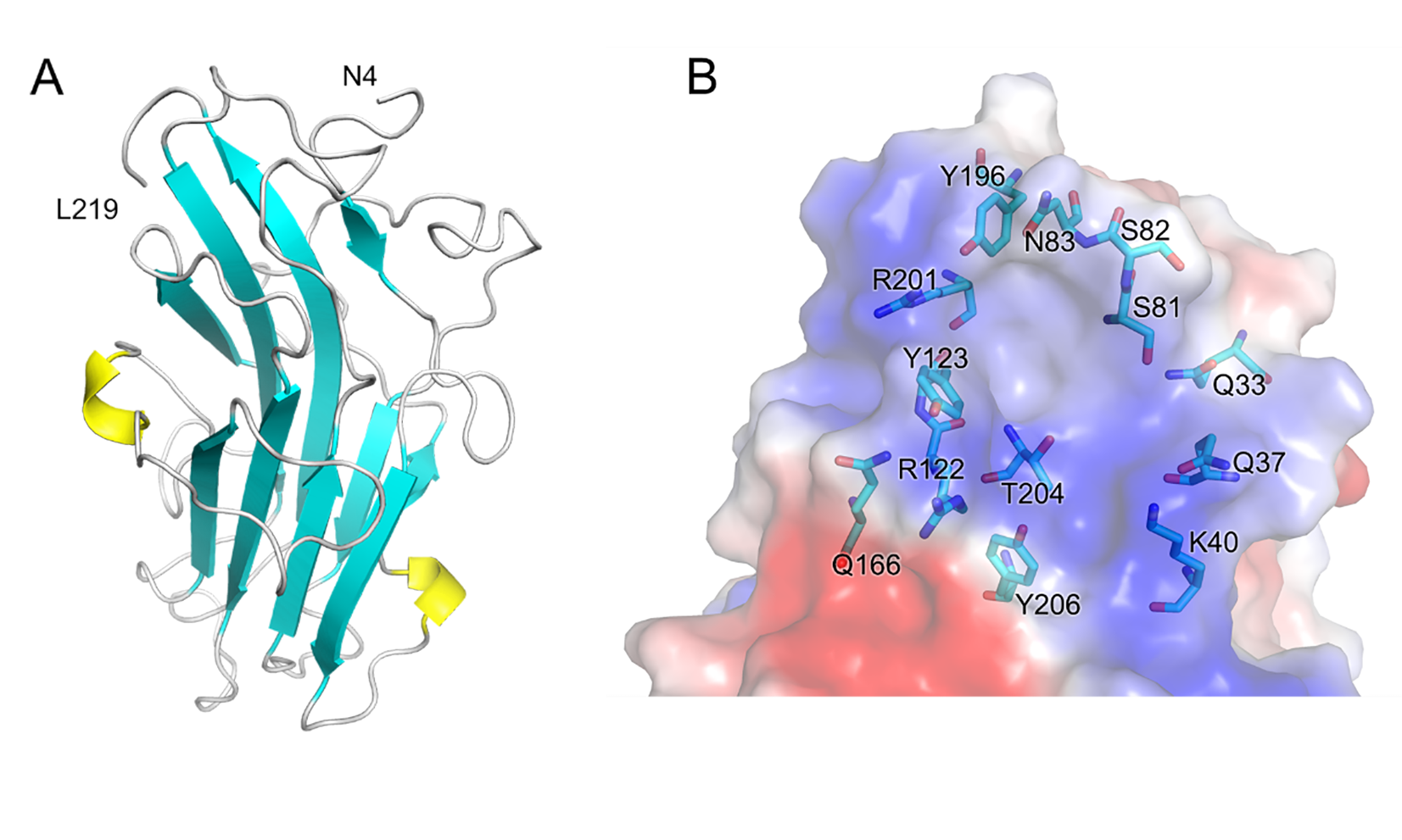
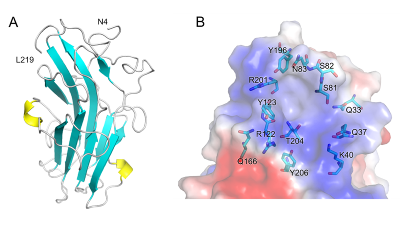
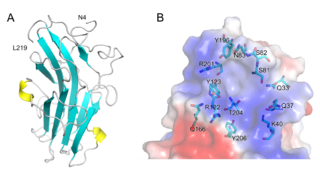
Structure of PhCBM100. (A) Schematic representation of PhCBM100 showing helices in yellow and strands in cyan. (B) Detailed view of the putative PhCBM100 binding site. The surface is colored by electrostatic potential, from blue (positive charge) to red (negative charge).
File history
Click on a date/time to view the file as it appeared at that time.
| Date/Time | Thumbnail | Dimensions | User | Comment | |
|---|---|---|---|---|---|
| current | 19:52, 4 January 2024 | | 1,653 × 991 (1.74 MB) | Guanchen Liu (talk | contribs) |
You cannot overwrite this file.
File usage
The following page uses this file: